Overview
A hepatobiliary iminodiacetic acid (HIDA) scan is an imaging procedure used to diagnose problems of the liver, gallbladder and bile ducts.
For a HIDA scan, also known as cholescintigraphy or hepatobiliary scintigraphy, a radioactive tracer is injected into a vein in the arm. The tracer travels through the bloodstream to the liver, where the bile-producing cells take it up. The tracer then travels with the bile into the gallbladder and through the bile ducts to the small intestine.
A nuclear medicine scanner, called a gamma camera, tracks the flow of the tracer from the liver into the gallbladder and small intestine and creates computer images.
Products & Services
Why it's done
Gallbladder and bile duct
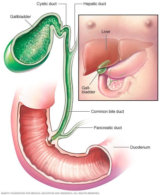
Gallbladder and bile duct
The gallbladder stores bile, a yellow-green fluid produced in the liver. Bile flows from your liver into your gallbladder, where it's held until needed during the digestion of food. When you eat, your gallbladder releases bile into the bile duct, where it's carried to the upper part of the small intestine, called the duodenum, to help break down fat in food.
A HIDA scan is most often done to evaluate the gallbladder. It's also used to look at the bile-excreting function of the liver and to track the flow of bile from the liver into the small intestine. A HIDA scan is often used with X-ray and ultrasound.
A HIDA scan might help in the diagnosis of several diseases and conditions, such as:
- Gallbladder inflammation, called cholecystitis.
- Bile duct obstruction.
- Congenital problems in the bile ducts, such as biliary atresia.
- Postoperative complications, such as bile leaks and fistulas.
- Assessment of liver transplant.
Your health care provider might use a HIDA scan as part of a test to measure the rate at which bile is released from your gallbladder, a process known as gallbladder ejection fraction.
Risks
A HIDA scan carries only a few risks. They include:
- Allergic reaction to medicines containing radioactive tracers used for the scan.
- Bruising at the injection site.
- Radiation exposure, which is small.
Tell your health care provider if there's a chance you could be pregnant or if you're breastfeeding. In most cases, nuclear medicine tests, such as the HIDA scan, aren't performed in pregnancy because of potential harm to the baby.
How you prepare
Food and medications
Your health care provider is likely to ask you:
- To fast for four hours before your HIDA scan. You might be allowed to drink clear liquids.
- To stop taking some medicines and supplements.
Clothing and personal items
You might be asked to:
- Change into a hospital gown.
- Leave jewelry and other metal accessories at home or remove them before the procedure.
What you can expect
Before the procedure
Your health care team will position you on a table, usually on your back, and inject the radioactive tracer into a vein in your arm. You might feel pressure or a cold sensation while the radioactive tracer is injected.
During the procedure
During the test, you may get an IV injection of the medicine sincalide (Kinevac), which makes your gallbladder contract and empty. Morphine sometimes is given during a HIDA scan to make the gallbladder easier to see.
A gamma camera is positioned over your abdomen to take pictures of the tracer as it moves through your body. This process takes about an hour, during which you'll need to remain still.
Tell your team if you become uncomfortable. You might be able to lessen the discomfort by taking deep breaths.
A specialist in medical imaging, called a radiologist, will watch a computer screen to see the progress of the radioactive tracer through your body. You might need more imaging within 24 hours if original images aren't good enough.
After the procedure
Most people can go about their day after the scan. The small amount of radioactive tracer will lose its reactivity or pass through your urine and stool over the next day or two. Drink plenty of water to help flush it out of your system.
Results
To make a diagnosis, your health care provider will consider your symptoms and other test results as well as the results of your HIDA scan.
Results of a HIDA scan include:
- Typical. The radioactive tracer moved freely with the bile from the liver into the gallbladder and small intestine.
- Slow movement of radioactive tracer. Slow movement of the tracer might indicate a blockage or obstruction, or a problem in liver function.
- No radioactive tracer seen in the gallbladder. Inability to see the radioactive tracer in the gallbladder might indicate acute inflammation, called acute cholecystitis.
- Low gallbladder ejection fraction. The amount of tracer leaving the gallbladder is low after medicine is given to make it empty. This might indicate chronic inflammation, known as chronic cholecystitis.
- Radioactive tracer detected in other areas. Radioactive tracer found outside of the biliary system might indicate a leak.
Your health care provider will discuss the results with you.