Overview
Parts of the immune system
Parts of the immune system
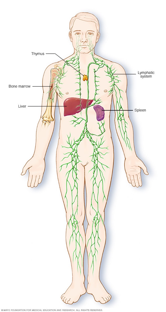
The lymphatic system is part of the body's immune system, which protects against infection and disease. The lymphatic system includes the spleen, thymus, lymph nodes and lymph channels, as well as the tonsils and adenoids.
Non-Hodgkin's lymphoma is a type of cancer that begins in your lymphatic system, which is part of the body's germ-fighting immune system. In non-Hodgkin's lymphoma, white blood cells called lymphocytes grow abnormally and can form growths (tumors) throughout the body.
Non-Hodgkin's lymphoma is a general category of lymphoma. There are many subtypes that fall in this category. Diffuse large B-cell lymphoma and follicular lymphoma are among the most common subtypes. The other general category of lymphoma is Hodgkin's lymphoma.
Advances in diagnosis and treatment of non-Hodgkin's lymphoma have helped improve the prognosis for people with this disease.
Products & Services
Types
Symptoms
Swollen lymph nodes
Swollen lymph nodes
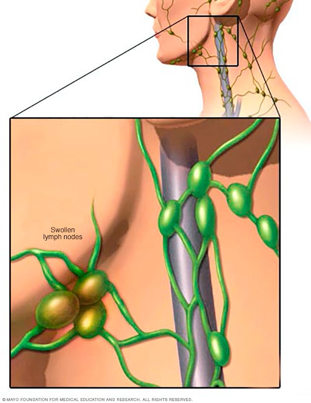
One of the most common places to find swollen lymph nodes is in the neck. The inset shows three swollen lymph nodes below the lower jaw.
Signs and symptoms of non-Hodgkin's lymphoma may include:
- Swollen lymph nodes in your neck, armpits or groin
- Abdominal pain or swelling
- Chest pain, coughing or trouble breathing
- Persistent fatigue
- Fever
- Night sweats
- Unexplained weight loss
When to see a doctor
Make an appointment with your doctor if you have any persistent signs and symptoms that worry you.
Causes
Lymph node clusters

Lymph node clusters
Lymph nodes are bean-sized collections of cells called lymphocytes. Hundreds of these nodes cluster throughout the lymphatic system, for example, near the knee, groin, neck and armpits. The nodes are connected by a network of lymphatic vessels.
In most instances, doctors don't know what causes non-Hodgkin's lymphoma. It begins when your body produces too many abnormal lymphocytes, which are a type of white blood cell.
Normally, lymphocytes go through a predictable life cycle. Old lymphocytes die, and your body creates new ones to replace them. In non-Hodgkin's lymphoma, your lymphocytes don't die, and your body keeps creating new ones. This oversupply of lymphocytes crowds into your lymph nodes, causing them to swell.
B cells and T cells
Non-Hodgkin's lymphoma most often begins in the:
- B cells. B cells are a type of lymphocyte that fights infection by producing antibodies to neutralize foreign invaders. Most non-Hodgkin's lymphoma arises from B cells. Subtypes of non-Hodgkin's lymphoma that involve B cells include diffuse large B-cell lymphoma, follicular lymphoma, mantle cell lymphoma and Burkitt lymphoma.
- T cells. T cells are a type of lymphocyte that's involved in killing foreign invaders directly. Non-Hodgkin's lymphoma occurs much less often in T cells. Subtypes of non-Hodgkin's lymphoma that involve T cells include peripheral T-cell lymphoma and cutaneous T-cell lymphoma.
Whether your non-Hodgkin's lymphoma arises from your B cells or T cells helps to determine your treatment options.
Where non-Hodgkin's lymphoma occurs
Non-Hodgkin's lymphoma generally involves the presence of cancerous lymphocytes in your lymph nodes. But the disease can also spread to other parts of your lymphatic system. These include the lymphatic vessels, tonsils, adenoids, spleen, thymus and bone marrow. Occasionally, non-Hodgkin's lymphoma involves organs outside of your lymphatic system.
Risk factors
Most people diagnosed with non-Hodgkin's lymphoma don't have any obvious risk factors. And many people who have risk factors for the disease never develop it.
Some factors that may increase the risk of non-Hodgkin's lymphoma include:
- Medications that suppress your immune system. If you've had an organ transplant and take medicines that control your immune system, you might have an increased risk of non-Hodgkin's lymphoma.
- Infection with certain viruses and bacteria. Certain viral and bacterial infections appear to increase the risk of non-Hodgkin's lymphoma. Viruses linked to this type of cancer include HIV and Epstein-Barr infection. Bacteria linked to non-Hodgkin's lymphoma include the ulcer-causing Helicobacter pylori.
- Chemicals. Certain chemicals, such as those used to kill insects and weeds, may increase your risk of developing non-Hodgkin's lymphoma. More research is needed to understand the possible link between pesticides and the development of non-Hodgkin's lymphoma.
- Older age. Non-Hodgkin's lymphoma can occur at any age, but the risk increases with age. It's most common in people 60 or over.