Overview
Spina bifida is a condition that occurs when the spine and spinal cord don't form properly. It's a type of neural tube defect. The neural tube is the structure in a developing embryo that later becomes the baby's brain and spinal cord and the tissues that enclose them.
Typically, the neural tube forms early in pregnancy and closes by the 28th day after conception. In babies with spina bifida, a portion of the neural tube doesn't close all the way. This affects the spinal cord and bones of the spine.
Spina bifida can range from being mild to causing serious disabilities. Symptoms depend on where on the spine the opening is located and how big it is. Symptoms also depend on whether the spinal cord and nerves are involved. When necessary, early treatment for spina bifida involves surgery. However, surgery doesn't always completely restore lost function.
Types
Spina bifida (myelomeningocele)
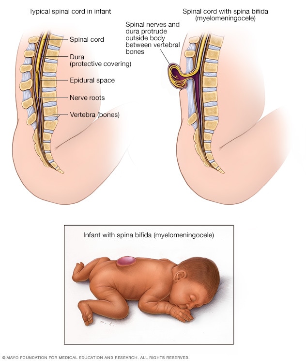
Spina bifida (myelomeningocele)
Myelomeningocele is the most serious type of spina bifida. The spinal cord's protective covering and the spinal nerves protrude at birth, forming a sac on the baby's back. The exposed nervous system may become infected, so prompt surgery is needed after birth.
Spina bifida occurs in different types: spina bifida occulta, myelomeningocele (my-uh-lo-muh-NING-go-seel) or the very rare type, meningocele (muh-NING-go-seel).
Spina bifida occulta
Occulta means hidden. Spina bifida occulta is the mildest and most common type. This type of spina bifida results in a small separation or gap in one or more of the bones of the spine, called vertebrae. Many people who have spina bifida occulta don't know they have it. It may be found during an imaging test such as an X-ray that is done for another reason.
Myelomeningocele
Myelomeningocele is the most serious type. It also is known as open spina bifida. The spinal canal is open along several vertebrae in the lower or middle back. Part of the spinal cord, including the spinal cord's protective covering and spinal nerves, push through this opening at birth, forming a sac on the baby's back. Tissues and nerves usually are exposed. This makes the baby prone to dangerous infections. This type also may cause loss of movement in the legs, and bladder and bowel dysfunction.
Meningocele
This is a rare type of spina bifida. In this type, a sac of spinal fluid bulges through an opening in the spine. No nerves are affected and the spinal cord isn't in the fluid sac. Babies with meningocele may have some minor trouble with functioning, including with the bladder and bowels.
Products & Services
Symptoms
Symptoms of spina bifida vary by type and from one person to another.
- Spina bifida occulta. Typically, there aren't any symptoms of spina bifida occulta because the spinal nerves aren't involved. But you can sometimes see symptoms on the newborn's skin above the small gap in the spine. You might see a tuft of hair, a small dimple or a birthmark. Sometimes, these skin marks can be symptoms of a spinal cord issue that can be found with MRI or a spinal ultrasound in a newborn.
- Meningocele. This type may affect bladder and bowel function.
- Myelomeningocele. In this most serious type of spina bifida, the spinal canal remains open along several vertebrae in the lower or middle back. The membranes and part of the spinal cord or nerves protrude at birth, forming a sac. Tissues and nerves usually are exposed, though sometimes skin covers the sac. Babies with this type of spina bifida may have trouble with bladder and bowel function. They also may experience weakness or lack of movement in the legs. Babies may have a buildup of fluid in the brain called hydrocephalus that can put pressure on brain tissue.
When to see a doctor
Typically, myelomeningocele is diagnosed before or right after birth, when medical care is available. Children diagnosed with this condition should be followed by a specialized team of healthcare professionals throughout their lives. Families can be educated on the different complications to watch for.
Children with spina bifida occulta typically don't have symptoms or complications. In these children, usually only routine pediatric care is needed.
Causes
The cause of spina bifida is not known. It's thought that a combination of genetic, nutritional and environmental risk factors causes the condition. This includes having a family history of neural tube defects and getting too little folate, also known as vitamin B-9, during pregnancy.
Risk factors
Spina bifida is more common among Hispanic people and white people. Also, female babies are affected more often than male babies. Although healthcare professionals and researchers don't know why spina bifida occurs, they have identified some risk factors:
- Too little folate in the pregnant person's body. Folate, the natural form of vitamin B-9, is important to the development of a healthy baby. Folic acid is the synthetic form that's found in supplements and fortified foods. If folate levels are too low, it's known as a deficiency. Folate deficiency increases the risk of spina bifida and other conditions that affect the neural tube.
-
Family history of neural tube defects. Having one child with a condition that affects the neural tube slightly increases the chance of having another baby with the same condition. The risk increases even more if two previous children have been affected by the condition.
Also, being born with a neural tube defect increases the chance of giving birth to a child with spina bifida. However, most babies with spina bifida are born to parents with no known family history of the condition.
- Some medicines. Taking anti-seizure medicines such as valproic acid during pregnancy increases the risk of having a baby with spina bifida. This might happen because the medicines interfere with the body's ability to use folate and folic acid.
- Diabetes. Having diabetes that is not well controlled before becoming pregnant increases the risk of having a baby with spina bifida.
- Obesity. Obesity at the time of pregnancy also is associated with an increased risk of spina bifida.
- Increased body temperature. Some evidence suggests that increased body temperature in the early weeks of pregnancy may increase the risk of spina bifida. A high core body temperature can be caused by a fever or by using a sauna or hot tub.
If you have known risk factors for spina bifida, talk with your healthcare professional. You may need a larger dose or prescription dose of folic acid, even before a pregnancy begins.
Also tell your healthcare professional about all of the medicines you take. If you plan ahead, some medicines can be adjusted to lower the potential risk of spina bifida.
Complications
Spina bifida may cause minimal symptoms or it can lead to more-serious physical conditions. Symptoms are affected by:
- The size and location of the opening in the spine.
- Whether skin covers the affected area.
- Which spinal nerves come out of the affected area of the spinal cord.
A number of complications may affect children with spina bifida. But not all children get all of these complications. Many complications can be treated.
- Walking and mobility problems. The nerves that control the leg muscles don't work properly below the area of the spinal cord that is affected. This can cause muscle weakness of the legs. Sometimes it can cause loss of movement, known as paralysis. Whether a child can walk depends on the location of the spine that's affected and the size of the opening in the neural tube. Ability to walk also depends on the care received before and after birth.
- Orthopedic complications. Children with myelomeningocele can have several complications in the legs and spine because of weak muscles. The complications depend on the location of the spine that's affected. Possible orthopedic issues may include:
- Curved spine, known as scoliosis.
- Changes that aren't typical, such as an inward-facing foot, known as clubfoot.
- Dislocation of the hip.
- Bone and joint conditions.
- Shortened, tight muscles, known as muscle contractures.
- Bowel and bladder symptoms. Nerves that supply the bladder and bowel usually don't work properly in children with myelomeningocele. As a result, they can't control their bowel or bladder. This is because the nerves that supply the bowel and bladder come from the lowest level of the spinal cord.
- Buildup of fluid in the brain, known as hydrocephalus. Babies born with myelomeningocele commonly have a buildup of fluid in the brain. This condition is known as hydrocephalus.
- Shunt malfunction. Shunts placed in the brain to treat hydrocephalus can stop working or become infected. Warning signs may vary. Some of the warning symptoms of a shunt that isn't working include:
- Headaches.
- Vomiting.
- Sleepiness.
- Irritability.
- Swelling or redness along the shunt.
- Confusion.
- Changes in the eyes, such as a fixed downward gaze.
- Trouble feeding.
- Seizures.
- Chiari malformation type 2. Chiari malformation (kee-AH-ree mal-for-MAY-shun) type 2 is common in children who have myelomeningocele. The brainstem is the lowest part of the brain above the spinal cord. In Chiari malformation type 2, the brainstem becomes longer and is lower than usual in the spinal canal or area of the neck. This can cause arm weakness and trouble with breathing and swallowing. Rarely, this condition can cause compression on this area of the brain. Surgery is needed to relieve the pressure.
- Infection in the tissues surrounding the brain, known as meningitis. Some babies with myelomeningocele may develop an infection in the tissues surrounding the brain. This potentially dangerous infection may cause brain injury.
- Tethered spinal cord. A tethered spinal cord is when the spinal nerves attach to tissue and stretch. This can happen with scar tissue that resulted from surgery. When a spinal cord is tethered, it's less able to grow as the child grows. It can lead to loss of muscle function in the legs, bowel or bladder. Surgery can limit the degree of disability.
- Sleep-disordered breathing. Sleep apnea or other sleep conditions can affect both children and adults with spina bifida, particularly those with myelomeningocele. They may need to be tested to see if they briefly stop breathing several times during sleep, known as sleep apnea. Getting treatment can improve health and quality of life.
- Skin problems. Children with spina bifida have less feeling in some parts of their bodies and may get wounds on their feet, legs, buttocks or backs. Blisters or sores can turn into deep wounds or foot infections that are hard to treat. Children with myelomeningocele have a higher risk of developing wounds in casts.
- Latex allergy. Children with spina bifida have a higher risk of an allergic reaction to natural rubber or latex products. A latex allergy may cause rash, sneezing, itching, watery eyes and a runny nose. It also can cause a dangerous condition in which the face and airways swell and make it hard to breathe, known as anaphylaxis. It's best to use latex-free gloves and equipment during childbirth and when caring for a child with spina bifida.
- Other complications. Other medical issues may arise as children with spina bifida get older. These complications may include urinary tract infections, gastrointestinal (GI) conditions and depression. Children with myelomeningocele may have learning disabilities. They may have trouble paying attention and learning reading and math.
Prevention
You can greatly reduce your risk of having a baby with spina bifida or other neural tube defects by taking folic acid supplements. Begin taking the supplements at least one month before becoming pregnant and continue taking them through the first trimester of pregnancy.
Get folic acid first
Having enough folic acid in your body by the early weeks of pregnancy is critical to prevent spina bifida. But many people don't discover that they're pregnant until this time. For this reason, experts recommend that all people of childbearing age take a supplement of 400 micrograms (mcg) of folic acid a day.
It's also helpful to eat foods that contain folate or have had folic acid added to them, known as fortification. Foods that are fortified with folic acid include:
- Enriched bread.
- Pasta.
- Rice.
- Some breakfast cereals.
Folic acid may be listed on food packages as folate, which is the natural form of folic acid found in foods.
Planning pregnancy
Adults who are planning pregnancy or who could become pregnant need to get 400 to 800 mcg of folic acid a day.
The body doesn't absorb folate as easily as it absorbs folic acid, and most people don't get the recommended amount of folate through diet alone. Vitamin supplements that include folic acid are necessary to prevent spina bifida. It's also possible that folic acid may help lower the risk of other conditions that may be present at birth. These conditions include a cleft lip, a cleft palate and some heart conditions.
It's also a good idea to eat a healthy diet that includes foods rich in folate or fortified with folic acid. This vitamin is present naturally in many foods, including:
- Beans and peas.
- Citrus fruits and juices.
- Egg yolks.
- Cow's milk.
- Avocados.
- Dark green vegetables, such as broccoli and spinach.
When higher doses are needed
If you have spina bifida or if you've previously given birth to a child with spina bifida, you need extra folic acid before you become pregnant. If you're taking anti-seizure medicines or you have diabetes, you also may benefit from a higher dose of this B vitamin. Check with your healthcare professional before taking additional folic acid supplements.
Dec. 19, 2023